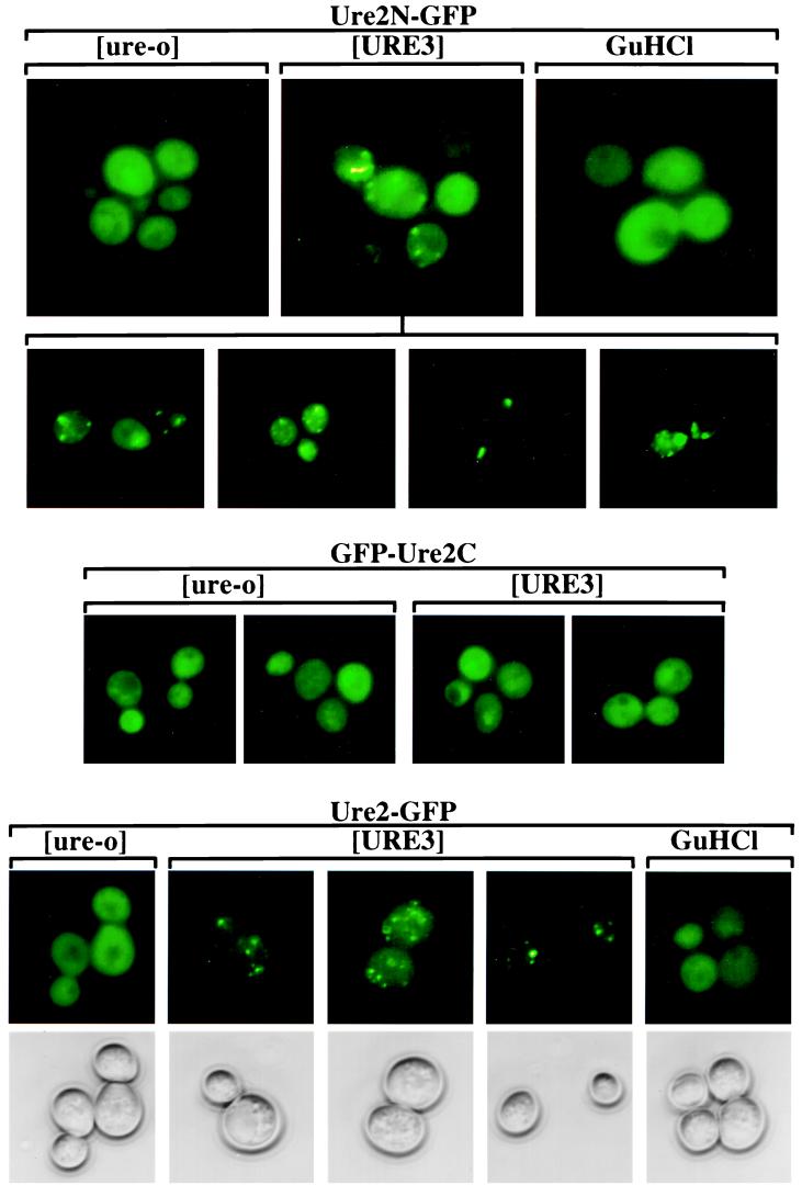
Figure 1.
Cellular distribution of Ure2-GFP fusion proteins. Into strains YHE64 (3686[URE3]) and 3686[ure-o] were introduced pVTG12 (CEN LEU2 PURE2 URE2N-GFP) (Top), pVTG13 (CEN LEU2 PURE2 GFP-URE2C) (Middle), and pH327 (CEN LEU2 PURE2 URE2-GFP) (Bottom). [ure-o], transformant of 3686[ure-o]; [URE3], transformant of YHE64 that retained [URE3]; GuHCl, transformant of YHE64 that was initially [URE3] but was cured by growth in the presence of 5 mM guanidine HCl. Phase contrast pictures of only the cells with pH327 (Bottom) are shown. The second row of pictures shows further examples of [URE3] cells with pVTG12.

