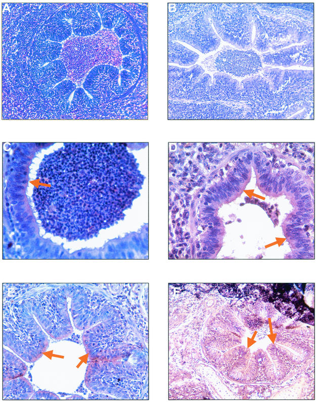
FIG. 4.
Histological and immunohistochemical findings as observed in the lungs of experimentally infected SPF pigs. (A) Peribronchiolar and perivascular accumulation of mononuclear cells with a mild hyperplasia of the bronchiolar epithelium (HE staining). (B to F) Immunoperoxidase staining by negative mouse serum (B) or by anti-P46 (C and D) and anti-P65c (E and F) MAbs of paraffin-embedded sections of lungs from experimentally infected pigs revealed staining patterns similar to that observed following immunofluorescence as indicated by arrows. Magnification, ×18 (A, B, E, and F) and ×36 (C and D).

