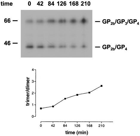
FIG. 4.
Formation of covalently linked GP2b/GP3/GP4 trimers. (Upper panel) The culture supernatant of [35S]cysteine-labeled EAV-infected cells was split into equal fractions and incubated at 39°C for the indicated time periods (in minutes). Next, the virions were dissolved by the addition of concentrated lysis buffer, and immunoprecipitations were performed with the serum directed against the GP2b protein. The numbers on the left are the molecular masses, in kilodaltons, of marker proteins analyzed in the same gel. (Lower panel) The ratio between the amounts of radiolabel incorporated into the GP2b/GP3/GP4 trimers and into the GP2b/GP4 dimers was determined by phosphorimager analysis and plotted against time.

