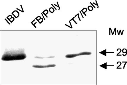
FIG. 2.
Comparative Western blot analysis of VP3 expressed in different systems. Extracts from IBDV-, VT7/Poly-, and FB/Poly-infected cells were subjected to SDS-PAGE and Western blot analysis with rabbit anti-VP3 serum, followed by addition of horseradish peroxidase-conjugated goat anti-rat immunoglobulin. Signal was detected by enhanced chemiluminescence. The position and molecular masses (in kilodaltons) of the immunoreactive VP3 bands are indicated.

