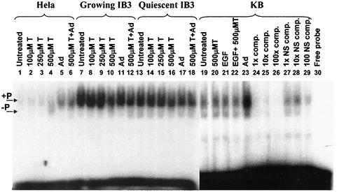
FIG. 4.
D-sequence EMSA analyses of HeLa, IB3, and KB cell extracts. Quiescent IB3 cells and exponentially growing HeLa, IB3, and KB cells were treated with increasing amounts of tyrphostin (T) for 2 h, and then the cells were harvested and WCE were prepared as described in Materials and Methods. Alternatively, cells were infected with adenovirus (Ad) for 36 h prior to preparation of the WCE. For adenovirus-infected tyrphostin-treated cells, the tyrphostin was added to the cultures for 2 h after the 36-h adenovirus infection. KB cells were also treated with 1 μg of EGF per ml for 24 h prior to WCE preparation. Equal amounts of protein were used to shift a radioactively end-labeled, single-stranded D-sequence oligonucleotide. KB cell extracts were also incubated with a 1-, 10-, and 100-fold excess of unlabeled competitor (comp.) D sequence or a nonspecific (NS) competitor oligonucleotide. The locations of the Tyr-phosphorylated ssDBP/DNA complex is indicated as +P. The Tyr-dephosphorylated form is indicated as −P on the left side of the figure.

