FIG. 6.
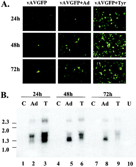
Northern analysis of mRNA from vAVgfp-transduced IB3 cells. Quiescent IB3 cells were left untreated, infected with adenovirus type 5 (Ad) at a multiplicity of infection of 10, or treated with tyrphostin and transduced with 100 particles per cell of vAVgfp. (A) At 24, 48, and 72 h later, expression was assessed by fluorescence microscopy prior to RNA isolation. (B) Northern analysis of RNA isolated from vector-transduced cells that were left untreated (lanes C), adenovirus-infected at a multiplicity of infection of 5 (lanes Ad), and treated with 500 μM tyrphostin (lanes T). A sample of RNA from untransduced cells (lanes U) was included. The locations of single-stranded DNA size markers (in kilobases) are shown on the left of the image.

