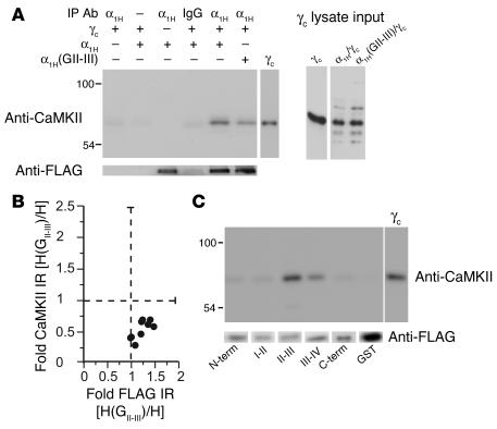
Figure 2. CaMKIIγC forms a signaling complex with α1H channels dependent on the II-III loop.
(A) Immunoblot of channel immunoprecipitates from HEK 293 cells transiently expressing FLAG-tagged α1H wild-type or α1H(GII-III) chimeric channels, most cotransfected with CaMKIIγC, using: goat IgG or anti-α1H for the immunoprecipitation, anti-FLAG for verification of channel immunoprecipitation, and anti-CaMKII for detection of CaMKII coassociation. Input lysates show expression level of CaMKIIγC with 0.25 pmol purified CaMKIIγC standard for comparison. (B) An analysis of 9 experiments. Integrated optical band densities of FLAG and CaMKII immunoreactivities (IR) in α1H(GII-III) channel immunoprecipitates expressed relative to immunoreactivities in wild type. (C) In vitro binding of purified CaMKIIγC (5 pmol/250 μl) to bead-bound channel intracellular domain–GST fusion proteins (2 pmol) of channel intracellular domains: N-terminus (N-term), I-II loop, II-III loop, III-IV loop, C-terminus. Shown are immunoblots of bound kinase detected by anti-CaMKII, with 0.5 pmol standard for comparison. Below is a GST blot for each fusion protein indicating nearly equal loading.

