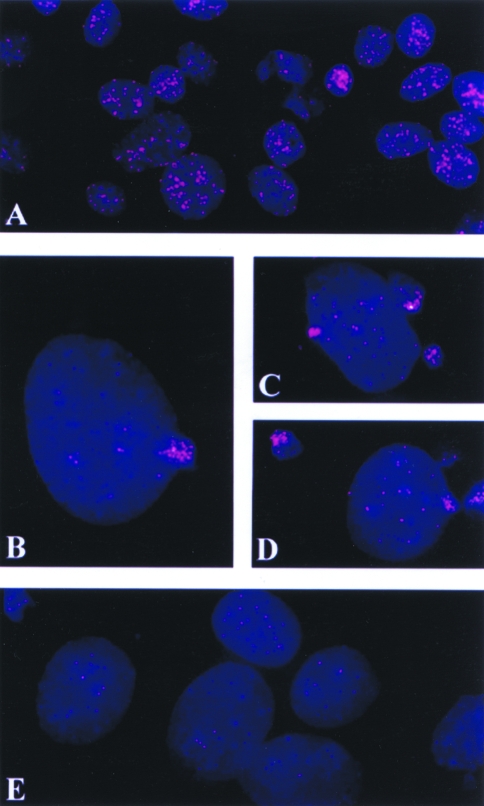
Figure 4.
FISH preparations of interphase cells of the K1735 clone X-21 cells, treated and untreated with ara-C. Cells are stained with DAPI for DNA (blue), and the telomeric DNA labeled with rhodamine (red). Untreated control cells showing a large quantity of telomeric DNA present (A). Telomeric DNA in bundles being extruded from nuclei of X-21 cells treated with ara-C for 24 and 48 hours (B, C, and D). Large nuclei from ara-C-treated cells showing reduced telomeric signals (E). All microphotographs are made at the same magnification.

