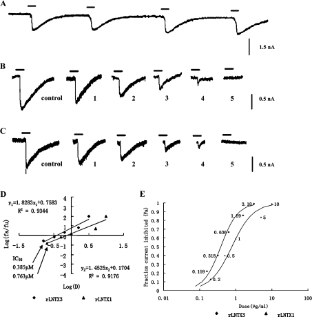
Figure 6. Inhibitory effects of rLNTX1 and rLNTX3 on nAChR-enriched skeletal myocytes.
Cells were held throughout the experiment at −70 mV. (A) Reproducible control currents excited by 80 μM nicotine. (B) Currents inhibited by rLNTX3 at a concentration of: 1, 0.159 μM; 2, 0.318 μM; 3, 0.636 μM; 4,1.59 μM; and 5, 3.18 μM. (C) Currents inhibited by rLNTX1 at a concentration of: 1, 0.20 μM; 2, 0.50 μM; 3, 1.00 μM; 4, 5.00 μM; and 5, 10.00 μM. (D) Analysis of the data by the median-effect plot. The median effect concentrations (IC50) of each peptide are marked. (E) Fraction of inhibited current to control current (fa) as functions of recombinant peptides at a series of concentrations (labelled beside the data points).

