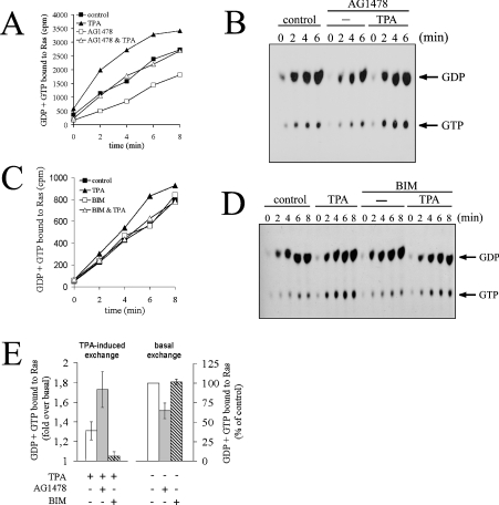
Figure 5. EGFR does not mediate GEF activation in response to PMA.
(A) Serum-starved COS-7 cells were pre-incubated with 200 nM AG1478 for 5 min at 37 °C or left untreated. Cells were challenged with 100 nM PMA (TPA) or left unstimulated, followed by permeabilization in the presence of [α-32P]GTP. Determination of guanine nucleotides associated with Ras was performed as described in the Experimental section. Note that PMA stimulation occurred 5 min prior to zero-time point as recorded in the plot. (B) Guanine nucleotides associated with Ras immunoprecipitates in (A) were separated by TLC. (C) The experiment was performed as described in (A), except that cells were pretreated with 500 nM BIM for 10 min. (D) Guanine nucleotides associated with Ras immunoprecipitates from the experiment shown in (C). (E) Quantification of nucleotide exchange for experiments shown in (A, C). Left panel: PMA-induced exchange. The amount of GDP + GTP bound to Ras 6 min after quenching the zero-time assay point (as recorded in A and C) was plotted as the fold increase in radioactivity associated with Ras in PMA-stimulated cells versus unstimulated cells for the various inhibitor treatments. Right panel: basal exchange rate. The amount of GDP + GTP bound to Ras in inhibitor-treated cells 6 min after quenching the zero-time assay point (as recorded in A and C) was plotted as percentage of radioactivity associated with Ras in untreated cells. Results shown represent the means and S.E. for at least three independent experiments.

