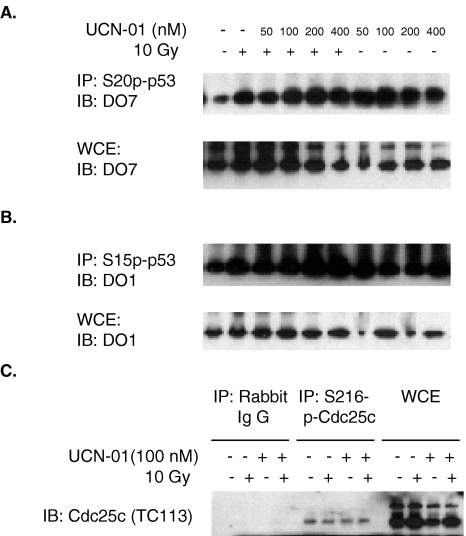
Figure 3.
Phosphorylation of p53 on S20 and S15 and phosphorylation of Cdc25C on S216 in HCT116/p53+/+ cells. After treatment with varying concentration of UCN-01 for 24 hours, cells were irradiated at 10 Gy and harvested 24 hours post-10 Gy for the whole-cell extract (WCE). (A) Phosphorylation of p53 on S20 in response to DNA damage and UCN-01 was performed by immunoprecipitation (IP) with antibody against S20p-p53 followed by an immunoblotting (IB) with p53 antibody, DO7. p53 protein levels were monitored by direct IB with DO7 without IP. (B) Phosphorylation of p53 on S15 in response to DNA damage and UCN-01 was performed by IP with antibody against S15p-p53 followed by an IB with p53 antibody, DO1. p53 protein levels were monitored by direct IB with DO1 without IP. (C) Phosphorylation of Cdc25C on S216 in response to DNA damage and UCN-01 was performed by IP with antibody against S216p-Cdc25C followed by an IB with Cdc25C antibody, TC113. Cdc25C protein levels were monitored by direct IB with TC113 without IP. Nonimmune rabbit serum was used as a control for the IP.

