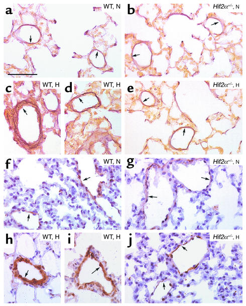
Figure 2.
(a–e) Hart’s elastin staining revealed the presence of vessels, located distally to the bronchi, at the level of alveoli and alveolar ducts, that contained only an IEL (or an IEL plus an incomplete EEL) (arrows) in lungs of normoxic (N) WT (a) and Hif2α+/– mice (b). Lungs of hypoxic (H) WT mice showed the presence of thick-walled vessels containing both an IEL and a complete EEL (arrows) (c and d), whereas no hypoxia-induced vascular remodeling occurred in Hif2α+/– mice (arrows) (e). (f–j) SMC α-actin staining shows the presence of partially muscularized peripheral vessels (arrows) in lungs of normoxic WT (f) and Hif2α+/– mice (g). Chronic hypoxia caused pulmonary vascular remodeling in WT mice, as revealed by the presence of fully muscularized vessels (arrows) (h and i), but not in Hif2α+/– mice (arrows) (j). Bar = 50 μm in all panels.

