
Figure 5.
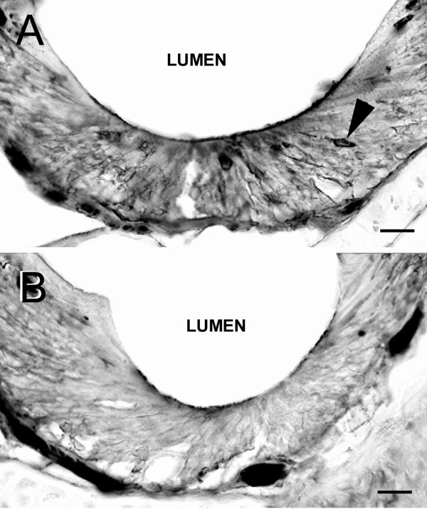
Micrographs of immunocytochemically labeled coronal sections of female P. shermani vomeronasal epithelium (VNE) demonstrating agmatine uptake by stimulation with PMF. (A) or a saline control solution (B). (A) Application of PMF resulted in the labeling of a moderate number of vomeronasal receptor neurons (arrow head) throughout all depths of the epithelial layer. Labeling of vomeronasal neurons by PMF was less intense than that by PRF. (B) Application of saline produced very few lightly labeled vomeronasal receptor neurons. Bars = 40 μm.