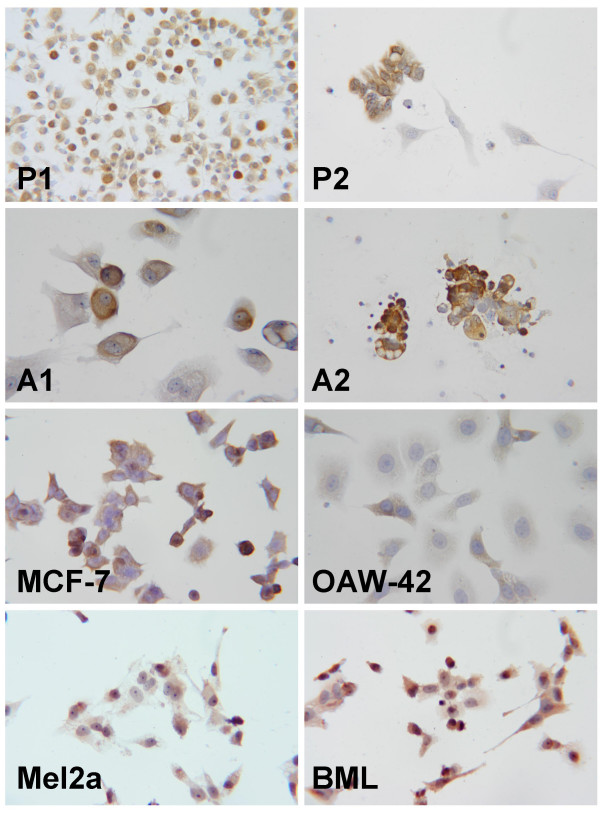
Figure 3.

Immunocytology of cells cultured from pleural effusions and malignant ascites (magnification 400×). a) Cells cultured from a malignant pleural effusion due to metastatic breast cancer. Staining of the cytoplasm for TRAP is seen. P1 b) Cells cultured from a malignant pleural effusion due to metastatic breast cancer. with marked expressison of TRAP in most of the cells. P2 c) Cells cultured from malignant ascites in a patient with ovarian cancer with obvious staining for TRAP. A1 d) Cells cultured from malignant ascites in a patient with ovarian cancer demonstrating staining for TRAP in the cytoplasm. A2 e) The commercially available cell line MCF 7 with staining for TRAP. f) The established cell line OAW42 also shows staining for TRAP. g) The malignant melanoma cell line Mel2A with moderate but unequivocal staining for TRAP. h) Malignant melanoma cell line BML with staining for TRAP.
