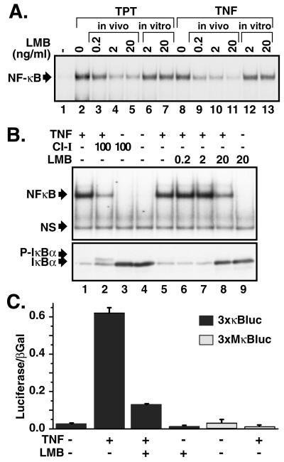
Figure 1.
LMB inhibits signal-inducible IκBα degradation and NF-κB activation. (A) PC-3 cells were treated with topotecan (TPT; 30 μM for 60 min; lanes 2–5) or TNFα (10 ng/ml for 15 min; lanes 8–11) in the presence or absence of various doses of LMB pretreatment for 30 min. Nuclear extracts were analyzed by EMSA by using an Igκ-κB probe. LMB was also added directly to nuclear extracts (as in lanes 2 and 8) and processed as described above (lanes 6, 7, 12, and 13). A section of the autoradiogram containing NF-κB complexes is shown. (B) HeLa cells were pretreated with CI-I (100 μM for 30 min; lanes 2 and 3) or LMB (ng/ml for 30 min; lanes 6–9) and then treated with TNFα (10 ng/ml for 15 min; lanes 1, 2, and 5–8). Nuclear extracts were analyzed by EMSA as described above (Upper). NS refers to a nonspecific band. Total cell extracts from parallel cell samples described in B were analyzed by Western blotting with IκBα antibody. P-IκBα refers to the position of the phosphorylated IκBα (Lower). (C) HEK293 cells were transiently transfected with a NF-κB-dependent reporter plasmid (3xκB-Luc or 3xMκB-Luc for control) and an internal control for transfection efficiency (CMV-β-Gal). At 36 h after transfection, cells were treated with or without TNFα in the presence or absence of LMB treatment. Cell extracts were analyzed for luciferase and β-galactosidase activities. Error bars are SD.

