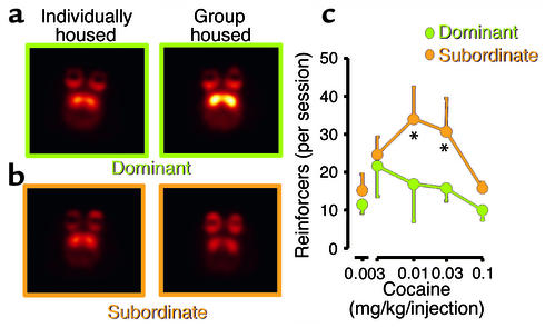
Figure 4.
Images of axial sections obtained with PET, showing DA D2 receptors in nonhuman primates that were initially tested while housed in separate cages and then retested after being housed in a group. Animals that became dominant when placed in a group (a) showed increased numbers of DA D2 receptors in striatum, whereas subordinate animals (b) did not. (c) The levels of cocaine administration in the subordinate and the dominant animals. Note the much lower intake of cocaine by dominant animals which possessed higher numbers of DA D2 receptors. The temperature scale was used to code the PET images; radiotracer concentration is displayed from higher to lower as yellow > red. Asterisks indicate significant differences in drug intake between groups. Adapted with permission from ref. 11.

