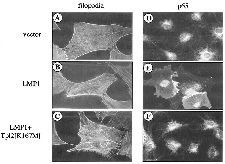
FIG. 4.
Tpl-2 is not a component of LMP1-induced Cdc42 activation. Serum-starved 3T3 fibroblasts were microinjected with pSG5-LMP1 (B and E) or control vector (A and D), in the presence or absence of myc-tagged Tpl-2[K167M] (C and F), as described in Materials and Methods. Phalloidin staining revealed formation of filopodia extensions in LMP1-expressing cells (B), which was not affected by the presence of kinase-inactive Tpl-2 (C). Note also the extensive formation of stress fibers in these microinjected cells. Parallel experiments using dominant negative Cdc42 verified inhibition of these LMP1-mediated cytoskeletal changes (data not shown), in agreement with a previous report (41). Transfection of the pSG5 vector alone had no effect on actin reorganization (A). Immunostaining for the p65 subunit of NF-κB (D, E, and F) revealed p65 translocation to the nucleus of LMP1-expressing cells (E) but predominantly cytoplasmic staining in control cells or cells coinjected with LMP1 and Tpl-2[K167M] (panels D and F, respectively).

