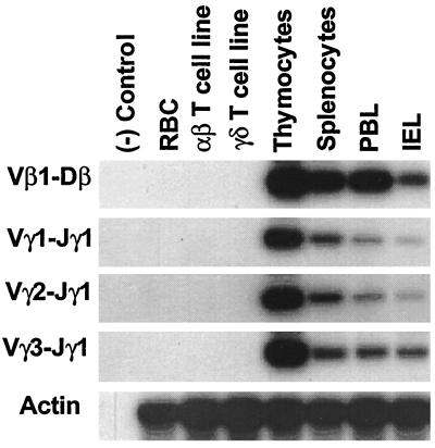
Figure 2.
Analysis of the tissue distribution of the deletion circles created by TCR V(D)J rearrangement. Lymphocytes were isolated from thymus, spleen, peripheral blood lymphocyte (PBL), and intestinal epithelial lymphocyte (IEL) compartments of a 4-week-old chicken and examined for extrachromosomal deletion circles by the PCR strategy illustrated in Fig. 1. PCR products separated in 1% agarose gels and transferred onto nitrocellulose membranes were hybridized with specific 32P-labeled internal oligonucleotide probes (Table 1) before membrane exposure to x-ray films for 12 hr.

