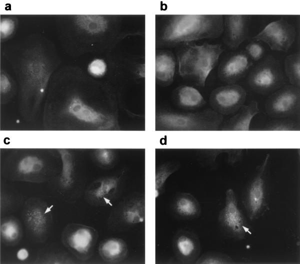
FIG. 5.
Detection of HAV antigen by immunofluorescence microscopy. Mock-infected (a and b) or HAV-infected (MOI = 1) (c and d) MO/MAC cultures were stained at 3 weeks p.i. with the HAV-specific antibody 7E7 and a fluorescein-labeled anti-mouse antibody. Cells were analyzed by fluorescence microscopy. Arrows indicate HAV antigen in the cytoplasm of infected cells. Magnification, ×400.

