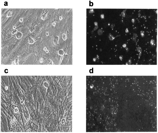
FIG. 7.
Detection of phagocytic cells in the stroma of HAV-infected LTBMCs. BMNC, isolated from patients undergoing hip surgery, were mock infected or infected with HAV (MOI = 1). For long-term culture, cultures were initiated in 12.5-cm2 flasks and incubated at 37°C in an atmosphere containing 5% CO2. The stromal layers of mock-infected (a and b) or HAV-infected (c and d) cultures were analyzed at 5 weeks p.i. for the presence of phagocytic cells, using the latex phagocytosis assay as described in Materials and Methods. Cells were analyzed by light (a and c) and fluorescence (b and d) microscopy. Magnification, ×200.

