FIG. 1.
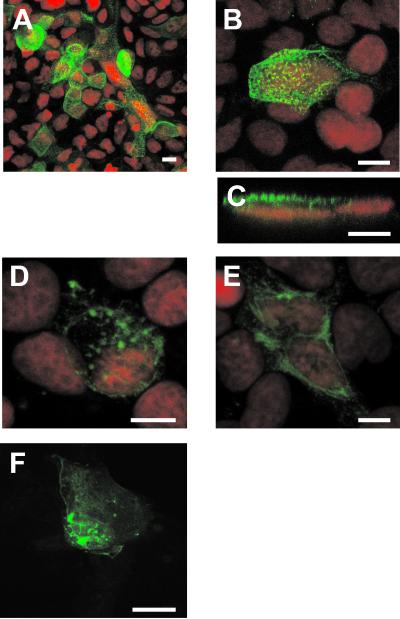
Early after rotavirus infection, VP4 is expressed at the apical membrane of Caco-2 cells and in discrete intracellular locations. Caco-2 cells (21 days old), grown on Transwell filters, were infected with the RF rotavirus strain at 10 PFU/cell (A to E). Six hours p.i., cells were fixed with 2% PFA and not permeabilized (A to C) or permeabilized (D and E). VP4 was detected using monoclonal antibody 7.7 and a FITC-labeled secondary anti-mouse IgG antibody. Nuclei were labeled with propidium iodide (red channel). (A) General representative view of the sum of the five most apical sections (1 μm deep each). (B) xy projection on a zoomed view. Ten 0.5-μm-thick sections were recorded from the top to the bottom and were projected on one plane to recover all the data available in the sample. (C) xz section obtained through an xz direct scanning procedure. (D and E) In permeabilized cells, it was possible to show that VP4 localized to discrete locations that evoked vesicular and tubular structures, which are illustrated as 1-μm-thick sections through the middle of the cells. (F) Cells were transiently transfected with VP4-GFP, as described in Materials and Methods, and directly observed with the confocal microscope within 48 h posttransfection. A sum of 15 sections, each 0.5 μm thick, is displayed. Bars = 10 μm.

