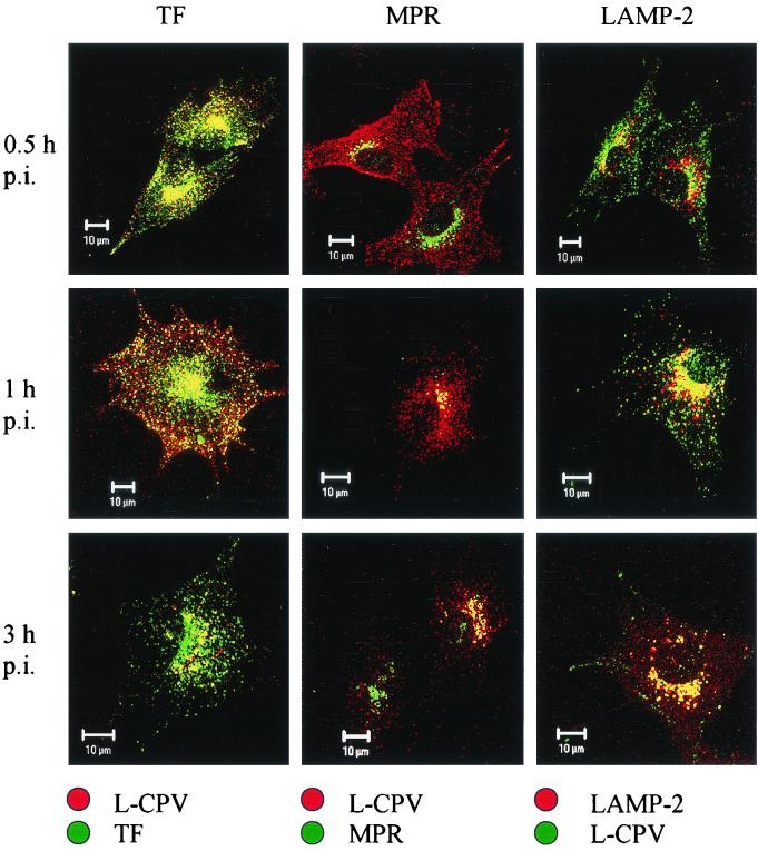
FIG. 10.
Colocalization of empty CPV capsids (L-CPV) and the organelle markers TF, MPR, and LAMP-2. The infected cells were fixed at time points p.i. indicated on the left. Left column: TF, green, L-CPV red. Middle column: MPR, green, L-CPV, red. Right column: L-CPV, green, LAMP-2, red. In all columns, the colocalization is yellow. Bars, 10 μm.

