
Figure 3.
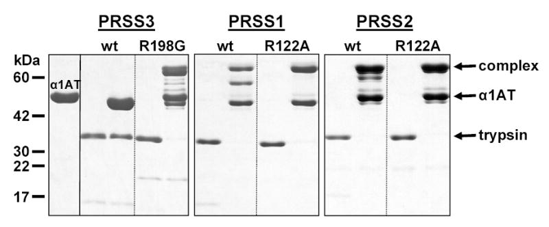
Covalent complex formation between wild-type α1-antitrypsin (α1AT) and human trypsins. Mesotrypsin (PRSS3), the R198G mesotrypsin mutant, cationic trypsin (PRSS1), the R122A cationic trypsin mutant, anionic trypsin (PRSS2), and the R122A anionic trypsin mutant were incubated at 1 μM concentration with or without 3 μM α1AT in 0.1 M Tris-HCl (pH 8.0), and 10 mM CaCl2, at 37 C° for 30 min. The 100 μL incubation mixes were precipitated with 10 % final concentration of trichloroacetic acid and subjected to reducing SDS-PAGE and Coomassie blue staining. The positions of the bands representing the covalent complex, the free α1AT and the free trypsins are indicated. See text for details on the bands migrating between the complex and free α1AT.