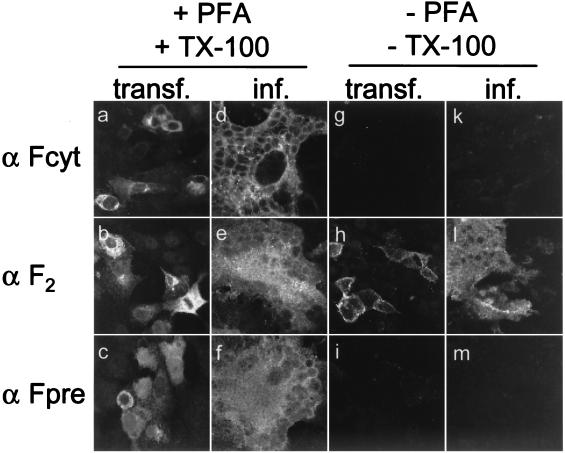
FIG. 5.
Immunofluorescence staining of Vero cells transfected with pCG-FOS or infected with CDVOS by using antibodies against different parts of the F protein. Cells were either fixed with paraformaldehyde (PFA) and permeabilized with Triton X-100 (TX-100) 48 h after transfection or infection with an MOI of 0.01 (a to f) or incubated unfixed with the primary antibody at 4°C before treatment with paraformaldehyde (g to m). Anti-Fcyt, anti-F2, or anti-Fpre rabbit antipeptide serum was used as the primary antibody.

