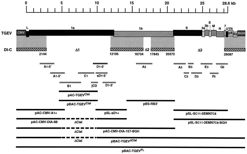
FIG. 1.
Assembly of the full-length TGEV cDNA. The assembly of a TGEV infectious cDNA (top) was started from the DI-C cDNA (discontinuous sequence indicated by the striped boxes). DI-C RNA has three deletions: Δ1 (10 kb), Δ2 (1.1 kb), and Δ3 (7.7 kb), which were filled in with newly synthesized RT-PCR products covering the rest of the viral genome (light grey bars). The sequence for the Δ1 deletion was amplified in six independent fragments: A1-5′, A1-3′, B1, C1, D1-5′, and D1-3′. The fragment D1-5′ (nt 9349 to 11173) could not be stably cloned. Nevertheless, the D1-5′ sequence was split into two overlapping clones (jCD [nt 8978 to 9758] and sD1-5′ [nt 9581 to 11173]) that were fully stable. Thus, the full-length cDNA was assembled in two large fragments that were finally joined by a ClaI9615 restriction site that split the toxic region. For that purpose, the ClaI4417 site was used as the other end of these fragments, while a third ClaI site, at position 18997, was removed by directed mutagenesis. A plasmid encoding the smallest fragment, including the viral sequence from nt 4310 to 9758 (pAC-TGEVClaI), was assembled by joining clones B1, C1, and jCD. Clones A1-5′ and A1-3′ were joined to the CMV immediate-early promoter (CMV) plus the 5′ end of the TGEV genome to make plasmid pAC-CMV-A1+. On the other hand, clones sD1-5′ and D1-3′ and the 5′ end of ORF 1b were joined to build plasmid pSL-sD1+, which was put together with pAC-CMV-A1+ to obtain a plasmid containing the 5′ third of the TGEV cDNA except for the ClaI deletion, called pAC-CMV-DIA-88. The Δ2 deletion was filled in with clone A2, which was joined to other ORF 1b sequences in plasmid pBS-RB3′. This was assembled with plasmid pAC-CMV-DIA-88 to obtain pAC-CMV-DIA-157-BGH. The Δ3 deletion was filled in with clones A3 to G3, from which plasmid pSL-Sc11-3EMN7c8 was assembled. The S gene was derived from the PUR46-C11 TGEV strain (Sc11) and genes 3, E, M, N, and 7 were from the PUR46-C8 strain (3EMN7c8), as well as the rest of the genome. The sequences necessary to make an accurate 3′ end, i.e., a synthetic poly(A) tail of 24 A residues (pA), the hepatitis delta virus ribozyme (Rz), and the bovine growth hormone termination and polyadenylation sequences (BGH) were added, yielding plasmid pSL-Sc11-3EMN7c8-BGH. Then a cDNA containing the whole TGEV genome except the ClaI4417-ClaI9615 fragment was assembled in the low-copy-number plasmid pACNR1180 (not shown). To improve the stability of the TGEV-derived cDNA sequences during their amplification in bacteria, the cDNA was transferred to pBeloBAC11, making plasmid pBAC-TGEVΔClaI. The ClaI4417-ClaI9615 restriction fragment was also cloned into pBeloBAC11, leading to plasmid pBAC-TGEVClaI. Insertion of the ClaI4417-ClaI9615 restriction fragment from pBAC-TGEVClaI into ClaI-linearized pBAC-TGEVΔClaI led to the full-length TGEV cDNA (pBAC-TGEVFL). Letters above the TGEV cDNA indicate viral genes. L, leader; UTR, untranslated sequences. Numbers below DI-C cDNA indicate positions of deletions with respect to the TGEV genome. ΔClaI, gap between ClaI4417 and ClaI9615 restriction sites.

