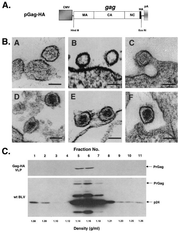
FIG. 1.
Overexpression of BLV PrGag leads to the production of VLPs. (A) Expression construct used for overexpression of PrGag. The gray box represents the cytomegalovirus promoter (CMV). The long white rectangular box represents the gag gene with the location of the corresponding MA, CA, and NC protein domains indicated. The influenza virus HA epitope tag is indicated by the thin black box. The bovine growth hormone polyadenylation signal (pA) is indicated. The locations of the HindIII and EcoRI restriction sites are indicated. (B) Visualization of VLPs by electron microscopy. Panels A to C show representative VLPs produced from COS-1 cells. The VLPs are immature and do not have core particles because the viral protease is absent. Panels D to F show representative wt virus particles produced from fetal lamb kidney cells chronically infected with BLV. The mature cores are readily visible. Bar, 100 nm. (C) Sucrose density gradient fractionation of VLPs. VLPs produced from COS-1 cells stably transfected with pGag-HA or wt BLV produced from fetal lamb kidney cells chronically infected with BLV were layered onto a sucrose gradient composed of 10, 20, 30, 40, 50, and 60% sucrose layers. The gradients were centrifuged, and 11 fractions were collected starting from the top of the centrifuge tubes. Fractionated samples were analyzed for BLV PrGag expression by Western blot analysis using either an anti-HA Ig (VLPs) or an anti-CA Ig (wt BLV). The locations of the 44-kDa Gag polyprotein (PrGag) and the 24-kDa capsid (p24) protein are indicated.

