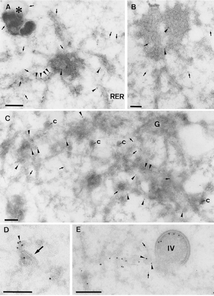
FIG. 8.
Double labeling of p16 and gp27 at 5 h postinfection. (A, B, and C) HeLa cells (A and B) or BHK cells (C) infected with MVA. All sections were double labeled with anti-p16 (10-nm-diameter gold) (arrowheads) and anti-gp27 (5-nm-diameter gold) (arrows). (A and B) Variable p16 labeling of the multimembrane vesicle that is connected to the rough ER (RER). These structures contain little to no labeling for gp27, while the membranes in continuity with them are clearly labeled for the cellular protein. In panel A gp27 but not p16 also labels a distinct dense vesicular structure (asterisk). (C) Relatively large amounts of label for both antigens in membranes adjacent to the Golgi complex are seen. The significant number of cop buds-vesicles (c) in continuity with these membranes indicates that they are on the cis side of the Golgi stack (G). (D) What we believe is the beginning of crescent formation (arrow) that is labeled with anti-p16 (arrowhead) is shown. (E) Double labeling with p16 (10-nm-diameter gold) (arrowheads) and gp27 (5-nm-diameter gold) (arrows), showing an IV, the inner membranes of which are labeled with anti-p16 and that is in continuity with tubular membranes that are labeled with both p16 and gp27. Bars, 200 nm.

