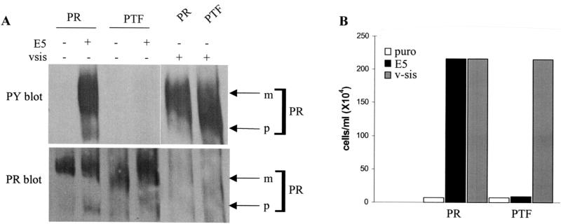
FIG. 6.
Biochemical and functional analysis of the triple substitution PTF mutant. Ba/F3 cells expressing the wild-type PDGFβR (PR) or the PTF mutant receptor with (+) or without (−) E5 or v-sis were established as described in Materials and Methods. (A) PDGFβR immunoprecipitates from cell extracts were immunoblotted with an antiphosphotyrosine antibody to assess receptor activation levels (top panel). The phosphotyrosine immunoblot was then stripped (see Materials and Methods) and reprobed with the PDGFβR antiserum to detect receptor expression levels (bottom panel). Each lane represents approximately 300 μg of extracted protein. Arrows on the right point to the mature (m) and precursor (p) forms of the receptors. (B) IL-3 independence assay of cells expressing the PTF mutant receptor. Ba/F3 cells expressing the wild-type PDGFβR (PR) or the PTF mutant receptor without (puro) or with E5 or v-sis were incubated in the absence of IL-3 for 11 days and then counted. The data are representative of multiple experiments listed in Table 1.

