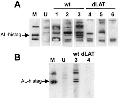
FIG. 6.
Detection of E. coli-expressed AL protein by sera from infected rabbits. (A) Total extract from E. coli expressing the AL-His tag fusion protein was run on a 15% Tricine gel with a single large loading well and transferred to a PVDF membrane. The membrane was cut into strips, and each strip was separately reacted with the indicated antibody. The strips were then reacted with horseradish peroxidase-conjugated secondary antibody for chemifluorescence. The arrow indicates the location of the E. coli-expressed AL-His tag fusion protein. Lane M, anti-His tag antibody as marker. Lane U, serum from an uninfected rabbit. Lanes 1 to 3, sera from three different rabbits infected with wild-type virus. Lanes 4 to 6, sera from three different rabbits infected with dLAT2903 (a LAT and AL-null mutant). (B) The E. coli-expressed AL-His tag protein was partially purified as described in Materials and Methods and processed as for panel A. Lane M, anti-His tag antibody as marker. Lane U, serum from an uninfected rabbit different from the serum in panel A. Lanes 3 and 4, the same rabbit sera as in lanes 3 and 4 in panel A. wt, wild type.

