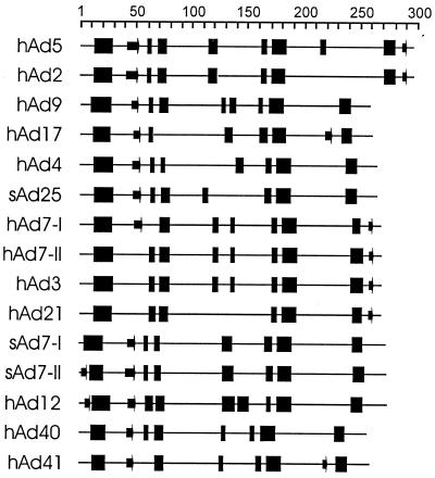
FIG. 6.
Secondary-structure predictions for the E1A proteins. Prediction of secondary structure for each of the indicated E1A proteins was performed using the PSIPRED program as described in Materials and Methods. Predicted α-helices and β-strands four or more residues in length are shown as blocks or arrows, respectively. The scale at the top indicates the amino acid positions within each E1A protein.

