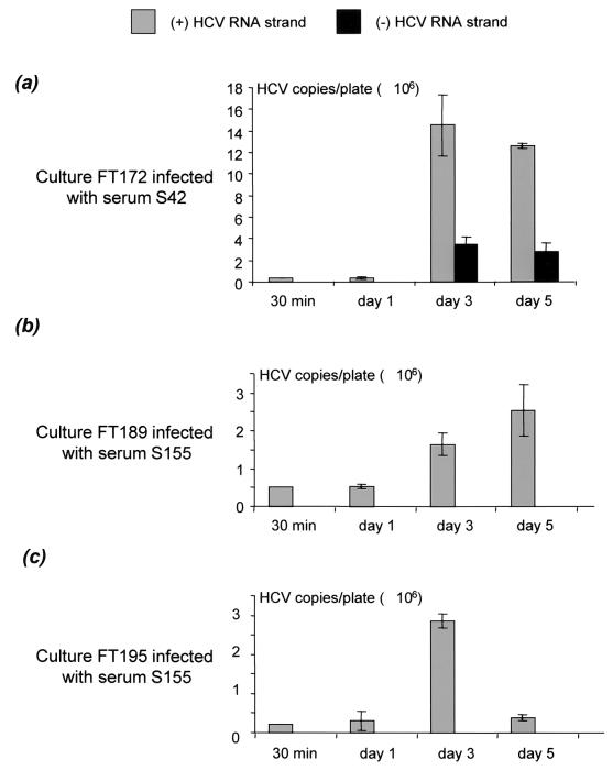
FIG. 3.
Accumulation of positive- and negative-strand HCV RNA in hepatocyte cultures FT172 (a), FT189 (b), and FT195 (c), infected with sera S42, S155, and S155, respectively, as measured by the quantitative LightCycler real-time RT-PCR assay. The hepatocyte cultures were infected 3 days after plating. The cells were harvested 30 min and 1, 3, and 5 days after infection for positive-strand (gray) and negative-strand (black) HCV RNA quantification. The amounts of HCV RNA strands are shown as means ± SEMs of three determinations, expressed in numbers of HCV RNA copies per 2 × 10⁶ cells, normalized to GAPDH mRNA. Similar results (not shown) were obtained with culture FT168 infected with serum S34.

