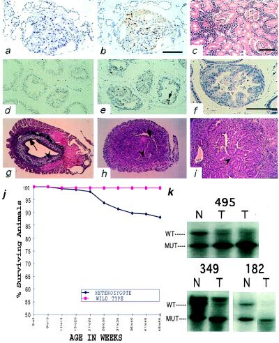
Figure 3.
Histological survey of tissues in Pten+/− mice. (a) Papillary thyroid carcinoma of 18-week-old Pten+/− male, hematoxylin/eosin (×400). (b) Anti-Ki67 stain of the same thyroid carcinoma. (c) A malignant lymphoma of the kidney of six-month-old Pten+/− male (case 87), hematoxylin/eosin (×200). (d and e) Immunohistochemical analysis of proliferation using anti-Ki67 antibody on prostates of +/+ (d) and +/− (e) mice. Arrow points to mitotic figure (×200). (f) Prostatic intraepithelial neoplasia found in 17-week-old Pten+/− male, hematoxylin/eosin (×400). (g–i) Mouse uteri. (g) Cross-section of the uterus of the 26-week-old wild-type mouse. Arrow points to the endometrium. Arrowhead points to the normal gland (×40). (h) Cross-section of the uterus of the 31-week-old heterozygous mouse. Arrow points to the endometrium. Arrowhead points to the atypical hyperplastic gland (×40). (i) Arrowhead points to a region of complex atypical hyperplasia in a 29-week-old heterozygous mouse (×100). (j) Decreased survival of the Pten heterozygous animals. (k) Loss of heterozygosity of Pten in tumors. Southern blots of normal-tumor pairs of three independent T-cell lymphomas analyzed with probe B. DNA was digested with SacI.

