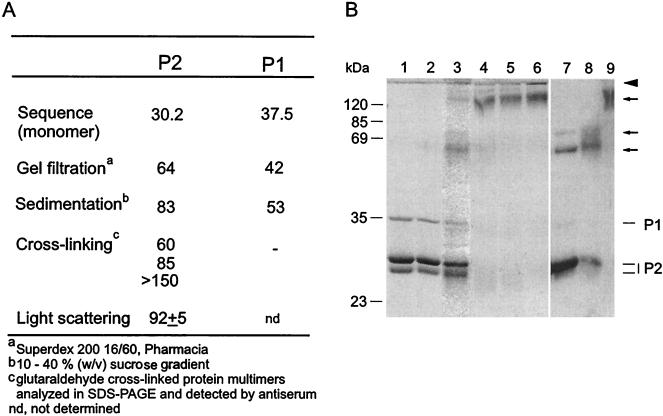
FIG. 4.
(A) Sizes of PM2 capsid proteins P1 and P2 (kilodaltons) obtained by different methods. (B) Cross-linking of proteins with glutaraldehyde. Lanes 1 to 6 show a Coomassie brilliant blue-stained polyacrylamide gel, and lanes 7 to 9 show a Western blot with anti-P2 serum. Non-cross-linked proteins P1 and P2 are shown in lane 1, and their positions are indicated on the right. Mixtures of proteins P1 and P2 (250 μg of protein/ml) were cross-linked with increasing glutaraldehyde concentrations (0.001% [vol/vol] [lane 2], 0.01% [lane 3], 0.05% [lane 4], 0.1% [lane 5], and 0.5% [lane 6]) as described in Materials and Methods. Multimeric forms detected by anti-P2 serum in samples cross-linked with 0.01% (lane 7), 0.05% (lane 8), and 0.1% (lane 9) glutaraldehyde are indicated by arrows. The boundary between the upper gel and the lower gel is indicated by an arrowhead. Numbers on the left indicate the molecular masses of the standard proteins.

