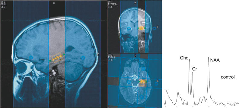
Figure 1. Single Voxel Image from the Left Hippocampus in an 11-y-Old Normal Male Child.
Blue areas are outer volume suppression bands that improve the accuracy of the hippocampal signal by suppressing surrounding lipid signal. On the right is a spectroscopy signal from a 11 year old male child, the spectrum demonstrating peaks from NAA, Cho, and Cr.

