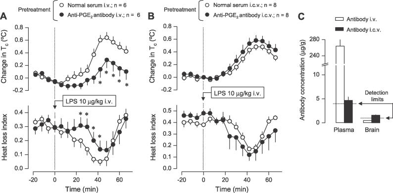
Figure 2. Circulating PGE2 Initiates LPS Fever in Rats: Direct Evidence.
(A) The effects of i.v. infusion (100 μl/kg/min, 120 min) of the anti-PGE2 antibody or normal serum 18 h before the experiment (pretreatment) on the Tc and heat loss index responses of rats injected (arrow) with LPS at thermoneutrality (30 °C).
(B) The effects of the i.c.v. infusion (2.7 μl/min, 15 min) of the same anti-PGE2 antibody or normal serum 18 h before the experiment (pretreatment) on the same responses. Note that the i.c.v. infusion was aimed at testing whether minute amounts of the antibody in the brain are sufficient to suppress LPS fever (and not at testing whether fever is altered by neutralization of PGE2 in the brain).
Change in Tc was calculated by subtracting the Tc value at a given point from that at the time of injection (time zero). In (A), the absolute Tcs at time zero were 38.2 ± 0.1 °C and 38.1 ± 0.2 °C for the groups treated with i.v. normal serum and antibody, respectively. In (B), the initial Tcs were 38.4 ± 0.1 °C and 38.2 ± 0.2 °C for the groups treated with i.c.v. normal serum and antibody, respectively.
(C) The levels of anti-PGE2 antibody in the blood plasma and whole brain of rats pretreated with i.v. or i.c.v. antibody. Blood samples and brains were collected immediately after the temperature responses were recorded, i.e., approximately 20 h after pretreatment with the antibody. Antibody levels (means ± SE) are expressed as microgram of neat antibody per gram of either plasma or brain tissue. The detection limit for each assay and the number of rats in each group (n) are indicated. An asterisk (*) indicates a significant difference from the group pretreated with normal serum (p < 0.05; two-way analysis of variance for repeated measures followed by the Tukey test).

