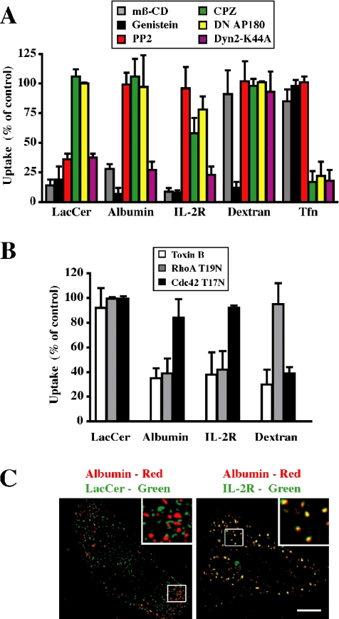
Figure 1.
Characterization of the endocytosis of various markers in CHO-K1 cells. (A) Cells were pretreated with mβ-CD, genistein, PP2, or CPZ or cotransfected with CFP-Nuc and DN AP180 or Dyn2 K44A constructs. Internalization (5 min at 37°C) of fluorescently labeled LacCer, albumin, dextran, or Tfn, relative to untreated cells (control) was quantified by image analysis. Internalization of IL-2R was followed after transfection of cells with IL-2R β (see Materials and Methods). (B) Cells were pretreated with toxin B or cotransfected with CFP-Nuc and RhoA T19N or Cdc42 T17N constructs, and internalization (5 min at 37°C) of the indicated marker, relative to that in nontransfected cells, was quantified by image analysis. Values in A and B represent means ± SD (n ≥ 30 from 3 independent experiments). (C) Colocalization of albumin with BODIPY-LacCer versus IL-2R in CHO cells. Cells, untransfected (left) or transfected with IL-2R β (right; see Materials and Methods), were coincubated with AF647-albumin and either BODIPY-LacCer or PE-mik-β3. After internalization for 1 min at 37°C, samples were acid stripped and back exchanged before microscopy. Separate images were acquired for each fluorophore, rendered in pseudocolor, and are presented as overlays. Insets show the boxed regions at higher magnification. Note that LacCer and albumin were not colocalized (presence of discrete green and red puncta) whereas LacCer and IL-2R overlapped extensively as shown by the yellow puncta. Bar, 10 μm.

