Figure 2.
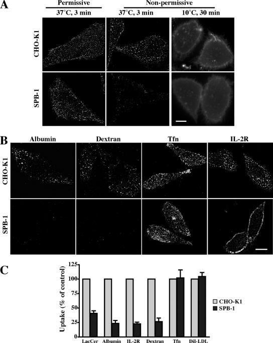
SL depletion selectively attenuates clathrin-independent endocytosis. (A) CHO-K1 or SPB-1 cells were cultured under permissive (F-12 medium containing 5% FBS at 33°C; left) or nonpermissive (Nutridoma-BO medium at 39°C; middle and right) conditions for 48 h. Cells were then incubated for 30 min at 10°C with 1 μM BODIPY-LacCer and immediately observed (right) or warmed for 3 min at 37°C and back exchanged (left, middle) before observation under the fluorescence microscope at green wavelengths. Similar effects were also observed after 5 and 10 min of internalization (Supplemental Figure 5B). (B) CHO-K1 or SPB-1 cells were cultured under nonpermissive conditions for 48 h. Internalization (5 min at 37°C) of the indicated markers was measured as in Figure 1. Bars, 10 μm. (C) Quantitative analysis of the uptake (5 min at 37°C) of the indicated markers in CHO-K1 and SPB-1 cells cultured under nonpermissive conditions. Results for SPB-1 cells are expressed as percentage of uptake measured in CHO-K1 cells. Values are the mean ± SD (n ≥ 50 cells from 3 independent experiments).

