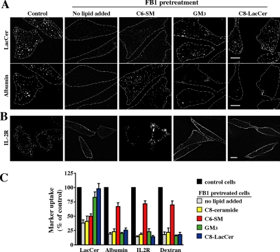
Figure 4.
Exogenous SLs restore marker uptake in FB1-treated cells. CHO-K1 cells were untreated (control) or pretreated with 20 μg/ml FB1 for 48 h. FB1-treated cells were then incubated with a 20 μM BSA complex of C6-SM, GM3, or C8-LacCer for 30 min at 10°C in HMEM. (A) Cells were then double labeled with BODIPY-LacCer and AF647-albumin for 30 min at 10°C, and the internalization (5 min at 37°C) was then measured as described in Figure 1. Dashed lines delineate cell periphery in each field. Bar, 10 μm. Note that the same fields of cells are shown for LacCer and albumin uptake. (B) Cells were incubated with IL-2R antibody under the same conditions as described in A. Fluorescent regions at the PM indicate noninternalized IL-2R antibody. Bar, 10 μm. (C) Uptake of BODIPY-LacCer, albumin, IL-2R, or dextran in FB1-treated cells after addition of the indicated exogenous SL. Results were quantified by image analysis and are expressed as a percentage of marker uptake in control cells. Values are the mean ± SD (n ≥ 50 cells for each condition).

