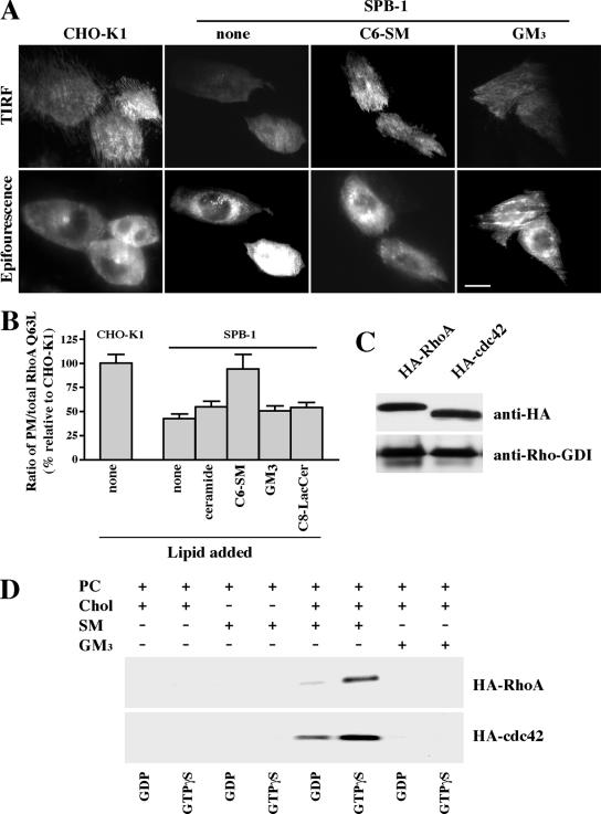
Figure 7.
Translocation of Rho-GTPases to membranes requires SM. (A andB) CHO-K1 and SPB-1 cells transfected with GFP-RhoA Q63L were cultured under nonpermissive conditions for 48 h and then serum starved. The SPB-1 cells were incubated for 30 min at 10°C ± 20 μM C6-SM/BSA or GM3/BSA in HMEM and then observed by TIRF or epifluorescence microscopy at 10°C. Bar, 10 μm. (B) PM-associated GFP-RhoA Q63L fluorescence was quantified relative to total cell fluorescence by image analysis as described in Figure 6E. PM/total cell fluorescence ratios calculated for SBP-1 cells ± lipids are expressed relative to the PM/total ratio in CHO-K1 cells (with no added lipid), which was set to 100. Values are the mean ± SD from ≥50 cells for each condition. (C) CHO-K1 cells were transiently transfected with HA-RhoA or HA-Cdc42. After 48 h, the HA-tagged proteins in the cell lysate were immunoprecipitated using immobilized anti-HA antibody matrix. The immunoprecipitates were analyzed by Western blotting using anti-HA or anti-Rho-GDI antibodies. (D) MLVs were prepared from DMPC/Chol (85/15, mol/mol); DMPC/SM (85/15, mol/mol), DMPC/SM/Chol (42.5/42.5/15, mol/mol/mol), or DMPC/GM3/Chol (42.5/42.5/15, mol/mol/mol) and subsequently incubated with HA-tagged RhoA or Cdc42 in the presence of GDP or GTPγS. After washing the MLVs, binding was determined by SDS-PAGE and Western blotting (see Materials and Methods).

