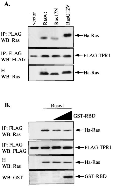
FIG. 7.
Interactions between Ras-TPR1 and Ras-Raf-1 RBD. (A) HeLa cells were transiently transfected with the FLAG-TPR1 construct either alone or in combination with expression vectors for the three forms of Ras. The FLAG-TPR1 protein was then immunoprecipitated (IP) from the cell lysates with an anti-FLAG agarose affinity gel. The Ras that was associated with FLAG-TPR1 was detected by Western blotting (WB) with an anti-Ras Ab (top panel). Ras17N exhibited reduced ability to bind TPR1. H, cell homogenates. (B) HeLa cells were transfected similarly as in panel A but with Raswt only. Cell lysates were prepared and preincubated for 30 min at 4°C with two different concentrations of purified GST-RBD derived from Raf-1, which were also present during immunoprecipitation of TPR1. The final reaction mixture contains 1.5 μg of cell lysates per μl and 6 and 27 ng of GST-RBD per μl. The Ras associated with FLAG-TPR1 was detected by Western blotting (top panel) and found to decrease with increasing concentrations of GST-RBD (bottom panel). Data shown are representative of at least three independent experiments.

