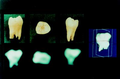
Figure 1.
Phosphorus-31 solid-state MRI of a human molar obtained by 3D projection imaging of 998 free induction decays at 2.0 T field strength. The three 2D images are planes of data selected from the full 3D data set. Alternatively, radiographic or dual energy x-ray absorptiometry-like projective views could have been easily shown as well. On the right is shown a solid body representation of the same data set, binary segmented by finding the surface containing all pixels above a threshold value and rendered by Gouraud shading.

