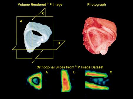
Figure 2.
Phosphorus-31 solid-state MRI of a section of fresh intact porcine bone cut from the tibial mid-shaft region, showing the solid body representation (Upper Left) and a photograph of the specimen (Upper Right). The presence of soft tissue does not interfere with the imaging process. The three 2D slices of the data set (Lower) correspond to the planes shown in the solid body rendition.

