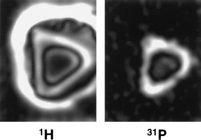
Figure 3.
Cross sections from 3D proton (Left) and phosphorus-31 (Right) solid-state MR images of an intact fresh specimen of midshaft porcine tibia. Note that cortical bone is bright in the proton image, as opposed to what it would be in conventional proton MRI. Muscle appears dark and subcutaneous fat is quite bright because of the spin-lattice relaxation times T1 of these tissues and the pulse repetition time used to acquire the proton image.

