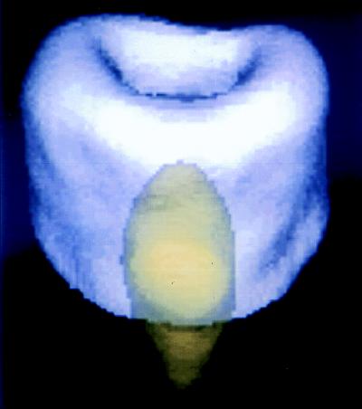
Figure 4.
Phosphorus-31 solid-state 3D projection reconstruction MR image of a section of the metaphyseal portion of porcine tibia containing a small capsule of synthetic hydroxyapatite powder to simulate a calcium phosphate-based prosthetic implant. The hydroxyapatite and the bone mineral, although chemically highly similar calcium phosphates, exhibit different MR spin lattice relaxation times T1. Two raw MR images, each with a different T1 sensitivity, were obtained and then appropriately combined on a pixel-by-pixel basis to yield chemically pure images of each material. The two images were then separately colorized and overlaid to produce the composite image. MR is unique among all 3D imaging methods applicable to solid materials, such as bone, ceramics, and polymers, because of its great sensitivity to chemical composition.

