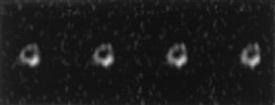
Figure 5.
Successive slices of a chemically sensitive phosphorus-31 solid-state 3D MR image of the mid-diaphyseal region of the femur of a live rabbit after surgical implantation of a cylinder of TCP, a prosthetic material commonly used for bone defect repair. The TCP mass projects partly out of the cortex (dense shell) of the femur. Enhancement of the TCP signal relative to the native bone mineral signal was achieved by using a large 90° RF pulse flip angle, which saturates the long T1 bone signal but not that of the short T1 implant material.

