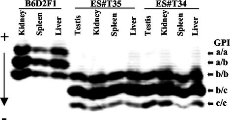
FIG. 1.
Analysis of GPI isoenzymes in tissue lysates of ES and control mice. Lysates of the indicated tissues of a B6D2F1 (GPI a/b) control mouse and two ES mice (ES#T35, ES#T34) derived from the ART4/12 ES cell line (GPI b/c) were separated by electrophoresis on a cellulose-acetate gel. The gel was stained for GPI enzyme activity and fixed. The run positions of the GPI homo- and heterodimers are indicated by arrows. The anode (+) and cathode (−) positions are indicated.

