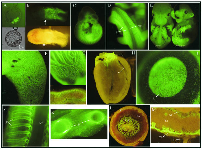
FIG. 2.
Expression patterns of αGFP in embryonic and adult tissues. (A) Expression in polar trophectoderm of E4.5 blastocysts. Bottom panel, visible light; top panel, UV. (B) Expression in extraembryonic ectoderm of an E6.5 embryo. Top panel, UV; bottom panel, visible light. Arrows denote the boundary of embryonic and extraembryonic tissues, and Reichert's membrane was not removed. (C) Whole-mount E10.5 embryo αGFP/+. (D) Magnification of the caudal somite. (E) E13.5 littermates. Left embryo, αGFP/+; right embryo, αGFP/αGFP. Expression in whole-mount E18.5 organs, lung (F), stomach (G, top portion), and transverse section of adult stomach in the glandular region (G, lower portion). (H) Sagittal section of adult heart. (I) Whole-mount adult eye. (J) Sagittal section of ribs and spinal cord of an E16.5 αGFP/+ embryo. (K) E18.5 αGFP/+ transverse section through ribs. Whole-mount (L) and transverse section (M) of an E14.5 αGFP/+ placenta visualized under fluorescence. ICM, inner cell mass; NT, neural tube; S, somites; V, ventricle; A, aorta; L, lens; R, ribs; P, perichondrium; La, labyrinth; ST, spongiotrophoblast layer; CA, chorioallantoic layer; U, umbilical cord.

