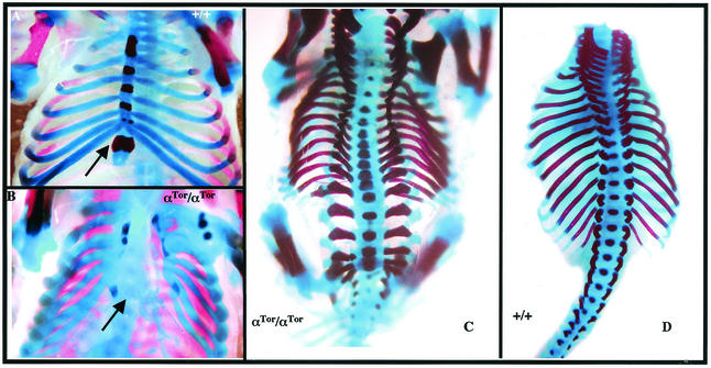
FIG. 5.
Skeletal defects in αTor mice. Skeletons of E16.5 embryos. (A) Wild-type ventral view of ribs and sternum. (B) αTor/αTor ventral view of rib cage. (C) αTor/αTor dorsal view of spinal column. (D) Wild-type dorsal view of spinal column with limbs removed. Arrows denote the positions of sternum.

