Figure 1.
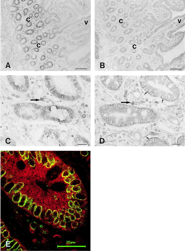
Immunohistochemistry of HFE protein and TfR in human duodenum. Immunoperoxidase staining of the HFE protein shows that the most intense signal is confined to the crypt enterocytes (A and C). The TfR shows wider distribution along the crypt-villus axis (B), but heavier staining at the basolateral surface of the crypt cells (D). Double immunostaining of the HFE protein and TfR demonstrates that both proteins are expressed in the same crypt enterocytes (E). The HFE protein shows a strong perinuclear staining pattern (C and E), whereas the TfR shows more diffuse intracellular as well as basolateral membrane-associated immunoreactions (small arrows in D). Leukocytes in the cellular lamina propria express both proteins (large arrows in C and D). The green and red colors in E indicate the HFE protein and TfR immunoreactions, respectively. Colocalization of HFE and TfR proteins is indicated by the yellow color. c, crypt region; v, villus. (Bars: A and B = 100 μm; C and D = 20 μm; and E = 25 μm.)

