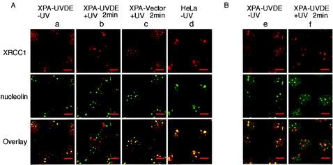
FIG. 2.
Nuclear distribution of XRCC1 and PARP-1 before and after local UV irradiation. (A) Double immunolabeling for nucleolin and XRCC1 in XPA-UVDE cells, XPA-Vector cells, and HeLa cells. Panels in column a are unirradiated XPA-UVDE cells, b and c are XPA-UVDE and XPA-Vector cells fixed at 2 min after local UV irradiation (20 J/m2), respectively, and panels in column d are unirradiated HeLa cells. Panels in the upper and middle rows correspond to XRCC1 (red) and nucleolin (green), respectively. Panels in the bottom row represent an overlay of the panels from the upper and middle rows. Colocalization of the two proteins appears yellow. (B) Double immunolabeling of XPA-UVDE cells for XRCC1 (red in the upper-row panels) and PARP-1 (green in middle-row panels); panels in the bottom row are the corresponding overlay; colocalization of the proteins appears yellow. Panels in column e are unirradiated cells, and those in f are cells at 2 min after local UV irradiation (20 J/m2). Bar, 10 μm.

